Scalable, real-time endogenous protein analysis
Biological Accuracy: Easily tag endogenous proteins while preserving natural function and localization
High-sensitivity: Consistently detect even low-abundance proteins for quantitative results in live or lysed cells
Speed & scalability: Get as many custom-edited, luminescence-verified cell lines as you require in as little as 7 weeks
Flexibility & confidence: Your desired cell lines and proteins, our guarantees and expertise to move your research forward
Custom Design. CRISPR Precision. Seamless Delivery.
HiBiT is an 11–amino acid tag that enables real-time, quantitative readout of endogenous protein expression without disrupting protein function or localization. When paired with Promega’s LgBiT and Nano-Glo® detection system, EditCo’s robust, HiBiT-tagged CRISPR knock-ins transform your cells into powerful biosensors — perfect for drug discovery, cell-based assays, and protein quantification.
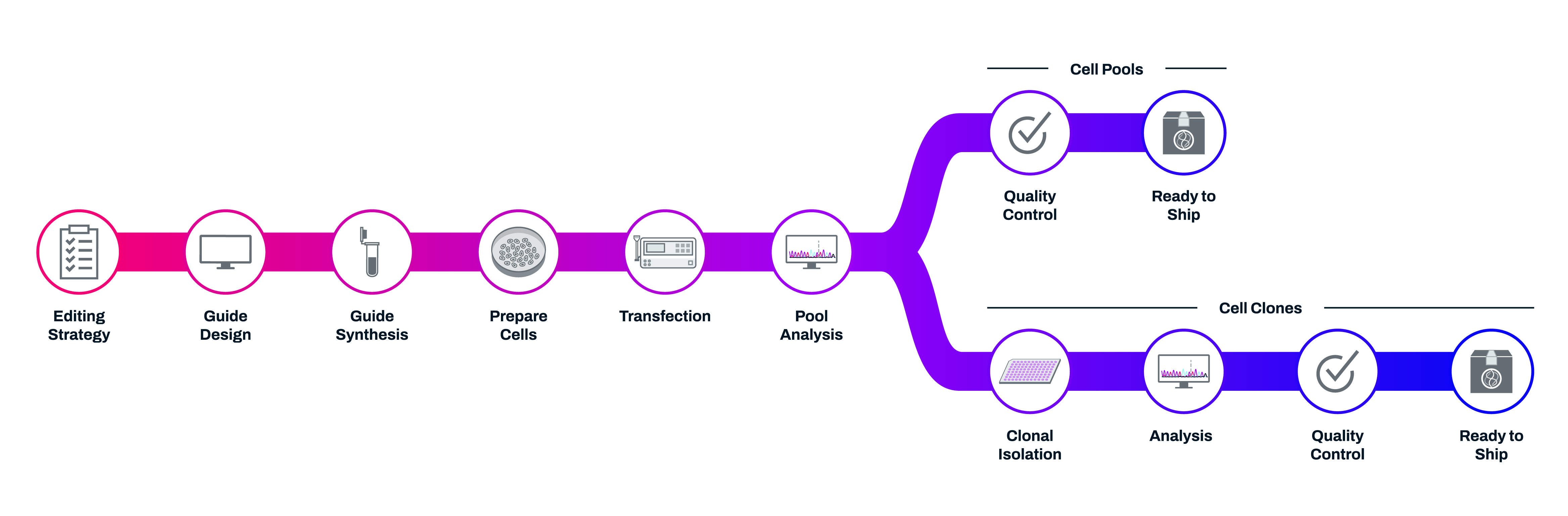
Our team of experts will design a precise knock-in strategy based on your gene of interest, cell line, and insertion location preference (C-terminal, N-terminal). Using RNP electroporation and synthetic guides, we maximize editing efficiency without introducing exogenous DNA elements
Cells available both clonally or as highly efficient pools with >70% average knock-in efficiency
Custom-Edited HiBiT-Tagged Cell Lines
Build or Buy? A Better Way to Tag Your Protein
EditCo provides across-the-board customization and confidence for both immortalized and iPS cells, regardless of scale, budget, gene popularity. While other HiBiT-tagged knock-in cell lines provide an easier solution than the internal burden of designing and editing tagged proteins yourself, they often have limitations or variable levels of support, flexibility, or guarantees.

Knock-in Cell Pools or Clones, Ready for Your Experiments
Deliverables
- Regular updates on your order's progress
- Edited cell pools or clones (2 vials with 500 thousand cells/vial)
- Mock-transfected cell pools (2 vials with 500 thousand cells/vial)
- Sequence of synthetic sgRNA used
- Primer sequences used for NGS sequencing
- NGS sequencing analysis report for each edited pool after expansion.
- Comprehensive QC report that includes mycoplasma test (positive/negative), passage number, and analysis for add-on QC
Features
Cell Source
- EditCo supplied
- Customer supplied
CRISPR Design
- Synthetic modified sgRNA (standard)
- Donor ssODN (standard)
Add-Ons
- QC: Luminescence
Sequencing deliverable note: For non-human/mouse cell types an alignment between the Sanger sequencing data of the edited pool or clone and the knock-in sequence will be provided.
Using CRISPR Knock-ins to Monitor Protein Abundance, Internalization, and Regulation
Determining Regulated Protein Abundance
Cellular protein levels shift in response to environmental signals or therapeutic intervention — and measuring those changes is essential for understanding biology and drug response. The rise of targeted protein degradation has unlocked a new modality for drug discovery, especially for previously “undruggable” targets.
By using CRISPR/Cas9 to insert a bioluminescent tag like HiBiT at the endogenous gene locus, researchers can quantify protein abundance with precision — no antibodies or overexpression required. These assays are highly sensitive, scalable, and compatible with both endpoint and live-cell formats, enabling real-time insight into native protein dynamics.

Endogenous tagging creates an improved assay window for HIF1A abundance assay. HeLa cells were transiently transfected with different amounts of CMV- or PGK-driven expression constructs for Hif1a-HiBiT; alternatively, HiBiT was tagged to the endogenous locus in HeLa cells using CRISPR/Cas9 gene editing. Cells were treated with a titration of 1,10-phenanthroline as indicated and protein abundance measured using the Nano-Glo® HiBiT Lytic Reagent.
(Source: https://www.promega.com/applications/small-molecule-drug-discovery/crispr-cas9-knock-in-tagging/)

Degradation kinetics of endogenous HiBiT-BRD4 following PROTAC treatment. HiBiT was inserted at the endogenous BRD4 locus in the HEK293 LgBiT Cell Line. Cells were treated with a titration of MZ1 in CO2-independent medium containing Nano-Glo® Endurazine™ Substrate. Luminescence measured in real time on the GloMax® Discover microplate reader.
(Source: https://www.promega.com/applications/small-molecule-drug-discovery/crispr-cas9-knock-in-tagging/)
Internalization of Cell Surface Receptors
Many cell surface receptors are internalized upon ligand binding as part of their natural signaling cycle. By inserting a HiBiT tag at the extracellular terminus of the receptor using CRISPR/Cas9, you can monitor internalization and recycling in real time — without relying on antibodies or overexpression systems.

Quantification of endogenous membrane receptor internalization following compound treatment. Pools of HeLa cells that were previously CRISPR-edited to insert HiBiT at the endogenous EGFR locus were treated with EGF as indicated. The Nano-Glo® HiBiT Extracellular Detection System was used to specifically detect HiBiT-EGFR expressed at the cell surface.
(Source: https://www.promega.com/applications/small-molecule-drug-discovery/crispr-cas9-knock-in-tagging/)
Monitoring Downstream Target Regulation
Upstream signaling events often influence the expression of downstream genes. With CRISPR/Cas9, you can insert a HiBiT tag at the endogenous locus of a downstream target and directly measure changes in protein expression. These live-cell assays enable real-time tracking of how pathway activity translates into functional protein output — all without overexpression or complex antibody workflows.
 Monitoring cMyc levels following degradation of upstream regulatory proteins. MV-4-11 cells, a model of acute myeloid leukemia, were edited using CRISPR/Cas9 to insert HiBiT at the endogenous cMyc loci and co-express LgBiT. Cellular cMyc gene expression levels are positively regulated by BET proteins and should be down-regulated after loss of inhibition or BET family proteins. Following treatment with BET protein degrader compounds dBET1 and MZ1, cMyc levels were monitored in real time. A decrease in BET protein levels resulted in decreased expression of the downstream cMyc target.
(Source: https://www.promega.com/applications/small-molecule-drug-discovery/crispr-cas9-knock-in-tagging/)
Resources
Scalable, real-time endogenous protein analysis made simple
Live-cell compatible: Track dynamic, real-time cellular processes without perturbing native protein function
High sensitivity & specificity: Accurate quantification of low-abundance targets with minimal background
Versatile applications: Adaptable for diverse experimental designs and research needs
Streamlined workflows: Optimized to reduce assay complexity and variability for reproducible high-content or high-throughput studies
Cells available both clonally or as highly efficient pools with >70% average knock-in efficiency
Explore More with HaloTag® Knock-ins
Multi-functional Tagging for Dynamic Biology
HaloTag® is a self-labeling protein tag that covalently binds to synthetic ligands, enabling precise control over how — and when — you visualize or manipulate a protein. By inserting HaloTag at the endogenous gene locus with CRISPR, EditCo gives you native expression and real-time control of your protein’s behavior, interactions, and location — without disrupting function.
From high-resolution microscopy to pull-down assays and degradation studies, HaloTag is a powerful tool for studying protein fate across diverse cellular environments.
Why Choose HaloTag?
- Live-cell compatible — tag proteins with fluorescent dyes for real-time imaging
- Covalent and customizable — bind to ligands for imaging, purification, or degradation
- Highly specific — minimal background and no cross-reactivity with native proteins
- Dual-function — combine detection and capture in the same cell line
- Protein fate control — pair with HaloPROTACs or dTAGs for conditional protein degradation
When to Use HaloTag (vs. HiBiT or NanoLuc)

Multiplex with Immunocytochemistry
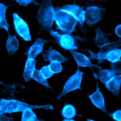
HaloTag® fusion proteins can be visualized alongside other cellular targets by combining fluorescent HaloTag ligands with immunocytochemistry. After labeling live cells with a HaloTag-compatible fluorophore, cells can be fixed and stained using an Anti-HaloTag polyclonal antibody paired with a spectrally distinct secondary antibody. This dual labeling approach helps validate in vivo ligand binding and ensures signal specificity. Because the antibody recognizes only the HaloTag protein, background staining is minimized, enabling clean, multiplexed imaging in fixed samples.

Colabeling of HaloTag®-p65 fusion protein with HaloTag® TMR ligand and the Anti-HaloTag® pAb. Cells were labeled with 5µM HaloTag® TMR Ligand for 15 minutes, gently washed and then incubated with 2µg/ml AlexaFluor® 488-conjugated anti-rabbit IgG (Invitrogen). Finally, cells were counterstained with DAPI (Vector Laboratories). Images were collected on the Olympus IX81 epi-fluorescent microscope with filter sets appropriate for rhodamine and AlexaFluor® 488. Panel A. Cytoplasmic (red) labeling of HEK293-p65-HT2 cells by HaloTag® TMR Ligand. Panel B. Cytoplasmic (green) labeling by Anti-HaloTag® pAb. Panel C. Colocalization of ligand and antibody binding activities. Panel D. Merger of red and green fluorescence with counterstaining of the nucleus by DAPI (blue). Arrows denote rare cells that show little or no expression of HaloTag®-p65.
Study Membrane Protein Localization and Trafficking
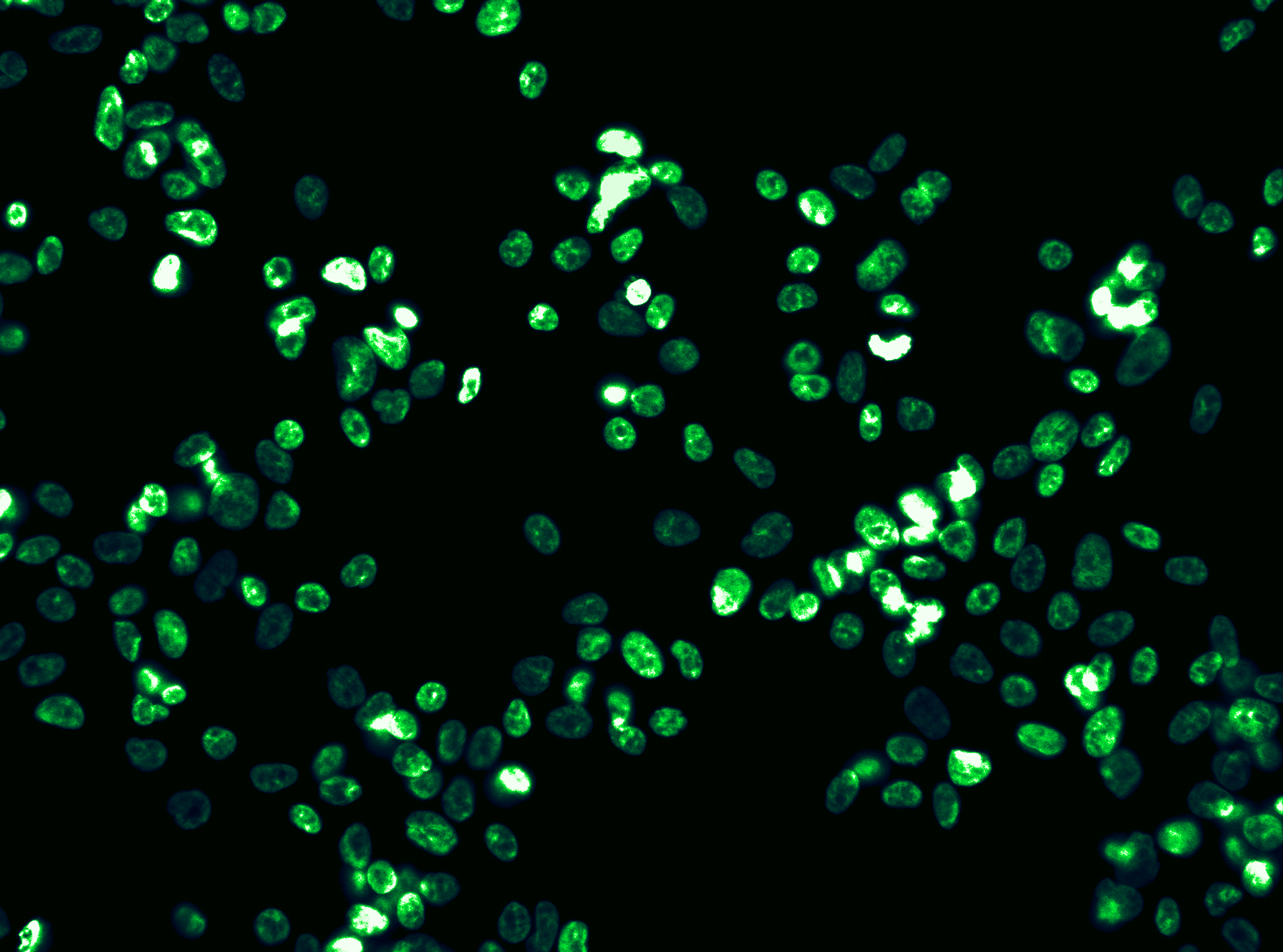
Track the localization and internalization of membrane-bound proteins by fusing them to HaloTag® and labeling with a fluorescent ligand that remains outside the cell. Since these ligands are cell-impermeant, they selectively bind only to surface-expressed proteins. The covalent interaction between the HaloTag protein and its ligand ensures the signal remains stable, even after fixation or under harsh experimental conditions. This allows you to confidently distinguish between surface-retained and internalized receptors, enabling detailed analysis of protein trafficking and membrane dynamics.

Live-cell labeling of cell surface HaloTag® fusion proteins. Panels A and B show HEK293 cells stably expressing HaloTag®-ECS (ExtraCellular Surface; comprised of a signal sequence and single transmembrane domain of b1-integrin) fusion protein labeled with HaloTag® Alexa Fluor® 488 Ligand and then imaged. Panels C and D show U2OS cells expressing HaloTag®-EDG1 (GPCR receptor) fusion labeled with Alexa Fluor® 660 Ligand and then imaged. Confocal images show fluorescence (Panels A and C) is restricted to the cell surface, also seen clearly in the panels with the DIC/fluorescence overlay (Panel B and D). Images were generated on an Olympus FV500 confocal microscope using the appropriate filter sets.
Achieve Superior Sensitivity with NanoLuc® Knock-ins
Ultra-Bright, ATP-Independent Luciferase for Sensitive, Scalable Detection
NanoLuc® luciferase is a next-generation, small (19.1 kDa) luciferase engineered for unparalleled luminescent performance. With >100-fold brightness compared to firefly or Renilla luciferase, NanoLuc enables high-sensitivity detection of low-abundance proteins and pathway activation — even in challenging biological systems.
When inserted at the endogenous locus via EditCo’s CRISPR knock-in workflow, NanoLuc becomes a native reporter, giving you a clean, quantitative signal without overexpression artifacts or the need for complex detection reagents.
Why Choose NanoLuc?
- Exceptional Brightness — delivers ultra-sensitive detection, even in low-expression systems
- Stable, Glow-Type Kinetics — ideal for endpoint or kinetic assays
- ATP-Independent Chemistry — less affected by cell health or energy status
- Small Size, Low Interference — preserves native protein function and dynamics
- Flexible Formats — supports secreted, intracellular, or split-luciferase (NanoBiT) designs

Resources
Integrating our core CRISPR expertise, high-quality reagents, and automated processes, we deliver the best edited cell-based models at any scale.

Use cases diam faucibus donec pretium leo risus commodo sit. Phasellus a sit.


Quisque amet eu viverra aliquam sed. Montes nulla nisi sit vel urna leo. Nisi donec consectetur bibendum tortor. Gravida arcu lorem venenatis nulla sodales quis sed.
Congue fermentum eros leo commodo. Integer vitae vitae ac amet. Vitae sed lectus vel lacus adipiscing nulla facilisis mauris porttitor. Viverra elementum odio vitae id.

Viverra aliquam volutpat interdum quisque vel mattis. Malesuada aliquam egestas pharetra a tempus ullamcorper egestas.
Nibh luctus pulvinar sagittis faucibus commodo vitae purus. Ullamcorper dictum.
Congue fermentum eros leo commodo. Integer vitae vitae ac amet. Vitae sed lectus vel lacus adipiscing nulla facilisis mauris porttitor. Viverra elementum odio vitae id.
Congue fermentum eros leo commodo. Integer vitae vitae ac amet. Vitae sed lectus vel lacus adipiscing nulla facilisis mauris porttitor. Viverra elementum odio vitae id.
Congue fermentum eros leo commodo. Integer vitae vitae ac amet. Vitae sed lectus vel lacus adipiscing nulla facilisis mauris porttitor. Viverra elementum odio vitae id.
Lorem Ipsum Dolor Heading
Etiam sollicitudin, ipsum eu pulvinar rutrum, tellus ipsum laoreet sapien, quis venenatis ante odio sit amet eros. Duis arcu tortor, suscipit eget, imperdiet nec, imperdiet iaculis, ipsum. Maecenas nec odio et ante tincidunt tempus. Donec elit libero, sodales nec, volutpat a, suscipit non, turpis. Quisque id mi.