
CRISPR-Cas9 is revolutionizing cell engineering, opening up new research possibilities such as easier disease modeling, clearer functional gene pathway analysis, novel drug target screening, or creating tailored immune cell therapies. However, not all cell lines are easily edited and often cell lines, such as primary immune cells, risk having their downstream functionality affected by the genetic changes or the editing process itself. One major hurdle researchers face when working with CD8+ T cells (also known as Cytotoxic T Cells or CTL) is maintaining their cytotoxicity after editing. The EditCo Bio R&D team recognized this challenge when developing our T cell editing process and was determined to succeed where others often falter.
The Challenge: Conserving Full Functionality After Editing
CD8+ T cells, also known as "killer T cells" or "cytotoxic T cells," are crucial for immune defense, as they identify and eliminate infected or cancerous cells. When CRISPR-Cas9 is used to knock out specific genes in these cells, the gene-editing process can cause stress that negatively affects their function. This impairment may result from the stress induced by the transfection methods or from the conditions during the recovery phase after editing.
However, preserving the functionality of CD8+ T cells is vital for obtaining reliable, reproducible, and meaningful results in any assays downstream from the editing process. These assays often aim to assess immune responses or the effects of specific genetic modifications. Impaired T cell functionality can compromise both the quality of the data and the validity of conclusions drawn from the experiments.
The Solution: Engineered Cells, Stronger Functionality
Video 1. Edited T Cells are seen killing a target cell via a BiTE antibody-mediated “redirected tumor killing” assay. Bright Field: CD8+ T Cells. Green: CFSE labeled Raji (CD19+) Cells. Red: Dead cells stained with DRAQ7 membrane impermeable viability dye.
At EditCo Bio, we’ve developed proprietary protocols to ensure that our CRISPR-edited CD8+ T cells retain full functionality due to optimized editing conditions and enhanced recovery protocols. Specifically, the functionality of edited CD8+ T cells has been assessed through target-cell killing, cytokine secretion, and proliferation assays.
To evaluate the ability of CD8+ T cells to kill target cells, such as tumor cells, bispecific T-cell engagers (BiTE antibodies) can be utilized.
How BiTE Antibodies Work to Provide Proof of T cell Functionality
Bispecific T-cell Engager (BiTE) antibodies are engineered immunotherapeutic molecules designed to bind simultaneously to two targets (Figure 1). These monoclonal antibodies have been widely tested in clinical trials and are already used for treating certain cancers.
In the case of the clinically approved blinatumomab (brand name BlincytoⓇ), one arm of the BiTE antibody binds to CD3e, a molecule present in all T cells, while the other arm targets CD19, a surface antigen found on cancerous B cells. This dual-binding mechanism effectively redirects T cells to target and kill cancerous B cells.
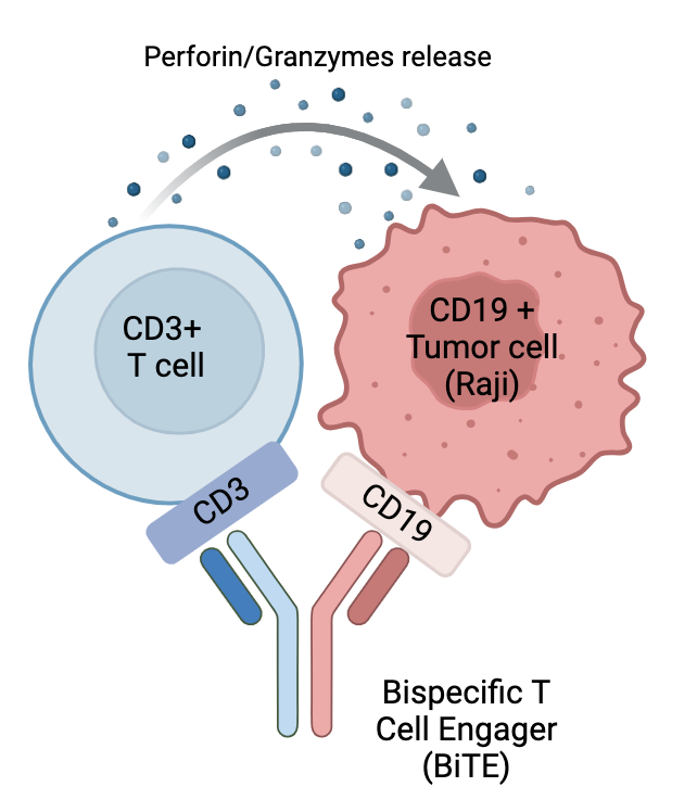
Figure 1. BiTE antibody schematic. This diagram illustrates the structure and function of a BiTE synthetic monoclonal antibody. One end binds to CD3e on CD8+ T cells, activating them, while the other end binds to CD19 on cancer cells (like Raji cells). This forms a bridge between the two cells, bringing them into proximity. The activated T cell then releases cytotoxic granules to destroy the cancer cell. This process is also known as "redirected tumor killing". (Figure created with Biorender)
EditCo’s CD8+ T cells Maintain Cytotoxicity Post-Editing
To assess the impact of CRISPR editing on the functionality of CD8+ T cells, we conducted a "redirected tumor killing" experiment using a BiTE reagent, as previously described. If the edited CD8+ T cells retain their full cytolytic activity, they should be able to kill CD19-expressing target cells (e.g., Raji B cells) at significantly higher levels compared to CD19-negative targets (e.g., K562 cells).
In detail, T cells from Editco’s off-the-shelf healthy donors were either activated or not activated for three days followed by mock-transfection with only SpCas9 protein. To track target cells in live microscopy and flow cytometry, Raji and K562 were incubated with CFSE, a membrane binding dye which colors cells green. After two days in culture to allow full recovery from the editing event, effector CD8+ T cells were incubated overnight with CFSE-stained Raji targets or CFSE-stained K562 targets (negative control) at a 10:1 effector to target ratio in the presence of anti-CD19-anti-CD3 BiTE antibody. The killing activity was detected by two different methods: live microscopy (Movie 1 and Figure 2) and flow cytometry (Figure 3).

Figure 2. Snapshots of a T cell killing a target cell via a BiTE antibody. In image 2A a T cell approaches a live cancer Raji B cell stained with CFSE (shown in green) and briefly samples it before lysing it in image 2B and becoming stained with the viability dye DRAQ7 (shown in red).

Figure 3. Flow cytometry data showing the capacity of edited CD8+ T cells to lyse cancer cells expressing specific antigens. These dot plots show Effectors (CD8+ T cells; anti-CD19 antibody stained) and Targets (K562 or Raji; CFSE stained) after 16 hours of incubation with non-activated (Panel A) and activated (Panel B) Effectors. The CFSE-labeled target events are significantly reduced when CD19+ Raji targets are used compared to CD19- K562 targets.

Figure 4. Quantification of the capacity of edited CD8+ T cells to lyse cancer cells expressing specific antigens. These histograms show the same data in Figure 3 in numerical format averaged across three replicate wells. Using either non-activated (A) and activated effectors (B), CD19 + Raji cells are killed far more efficiently ( 87.8% and 98.9%) compared to CD19 - K562 cells (53.5% and 46.8%).
Driving Innovation in Cell Therapy
These results showcase how EditCo Bio’s protocols to edit and recover CD8+ T cells preserve critical cell functionality, specifically their capacity to lyse cancer cells expressing specific antigens. At EditCo Bio, we don't just deliver edited cells—we deliver confidence. Whether you're investigating immune checkpoints, studying infectious diseases, or exploring novel targets, our CRISPR edited T cells are designed to perform under pressure.
If you're ready to bypass the common pitfalls of CRISPR editing and accelerate your research, we’re here to help. Connect with us to learn more about how our advanced cell engineering services can drive your research forward.
References
- Braun A, Gouni S, Pulles A, Strati P, Minnema MC, Budde LE. Bispecific Antibody Use in Patients With Lymphoma and Multiple Myeloma. Am Soc Clin Oncol Educ Book. 2024 Jun;44(3):e433516.
- Surowka M, Klein C. A pivotal decade for bispecific antibodies? MAbs. 2024 Jan-Dec;16(1):2321635.


